Neuron
Table of Content |
 (Unit of Nervous tissue-nerve cell)
Nerve cell is made up of cell body & cell process. (Dendron and axon = Neurites)
Cell body or cyton or soma or perikaryon:
It contains uninucleated cytoplasm.
Except centriole, all cell organelles are found in cytoplasm,
Centriole is absent or immaturely present in the nerve cell thus cell division is absent
Some other cell organelles like Nissl's granule and Neurofibril are also found in nerve cell.
Nissl's Granules
Endoplasmic reticulum 'coils around the ribosome & form granule 'like structure called as Nissl's granules or Tigroid body.
It is the centre of protein syntehsis,
Chemically - Ribonudeoprotein containing Iron.
Site - Cyton & dendron (Rod shape).
Many small fibrils are found in the cytoplasm called neurofibrils, these help in internal conduction in the cyton.
Cell Processes
Dendron: It is small cell process. It's fine branches are called dendrites. Some receptor's are found on the dendrites, so dendron receive the stimuli & produce centripetal (towards the cell body) conduction.
Axon: (Long process = Axon = Nerve fibre)
It is longest cell process of cyton, its diameter is uniform. It contain axoplasm.
Nissl's granules are absent in the axoplasm. Axoplasm of axon contains only neurofibrils and mitochondria).
Axon is covered by axolemma. Part of cyton where axon arises is called axon hillock.
The axon hilloick is the neuron's trigger zone, because it is the site where action potential are triggered.
The terminal end of axon is branched in button shape branches which are called as Telodendria.
More mitochondria are found in the telodendria which synthesize acetylcholine (Ach) with the help of choline acetyl transferase enzyme.
Ach is stored in the vesicles.
Axon is the functional part of nerve cell therefore term nerve fibre usually refer to axon.
Axon is covered by a layer of phospholipids (sphingomyelin) which is called as medulla or myelin sheath.
Medulla is covered by thin cell membrane which is called as neurilemma or sheath of schwann cells.
The neurilemma is composed of schwann cells.
Schwann cells take part in the deposition of myelin sheath (myelinogenesis).
Myelin sheath acts as insulator and prevent's leakage of ions.
Myelinogenesis in the Peripheral Nervous System (PNS)
In the peripheral nerves, myelinogenesis begins with the deposition of myelin sheath in concentric layer around the axon by schwann cells.
Myelin sheath is discontinuous around the axon.
These interruptions where axon is uncovered by myelin sheath are called node of Ranvier.
Myelinogenesis in the central nervous system (CNS):
Neurilemma or schwann cells are not present therefore myelinogenesis process occurs with the help of oligodendrocytes (Neuroglia).
Neurons in which myelin sheath is present are called medullated or myelinated neurons. In some nerve cells where myelin sheath is absent, they are called as non medullated or non myelinated neurons.
Collaterals of Axon:
These are small process or branches of axon. It's structure is similar to axon. It help in the conduction of nerve impulse.
Gray matter: It is composed of nerve cells. It consist of cytons & non medullated nerve fibres.
White matter: It contain myelinated nerve fibres
Types of Neurons
|
Unipolar |
Bipolar |
Multipolar |
|
Single process arises from cyton (1 Axon) e.g. Nervous system of embryo |
Two process arises n from cyton (1 Axon & 1 dendron e.g. Retina (Rod & cones) Olfactory epithelium |
Neuron which have one axon but many dendrons. e.g. Most of neurons of vertebrates. |
Apolar/Nonpolar Neuron: No definite dendron/ axon. Cell process are either absent or if present are not differentiated in axon and dendrons. Nerve impulse radiates in all directions. e.g. Hydra, Amacrine cell of Retina.
Pseudo unipolar: In this type, cell has only axon but a small process develop from axon which act as dendron.
eg. Dorsal root ganglia of spinal cord.
Active and passive ion movemen1ts across the cell surface of an axon. The movements are responsible for the generation of a negative/potential inside the axon. This is called the resting potential. Active transport takes place through the sodium/ potassium pump. Ion channels (proteins) allow the passive movement of ions down their electrochemical gradients.
The resting membrane potential in resting phase:
The potential difference (a charge) which exists across the cell surface membrane of nerve cells, is negative inside the cell with respect to the outside. The membrane is said to be polarised. The potential difference across the membrane at rest is called the resting membrane potential and this is about 70 mV (the negative sign indicates that inside the cell is negative with respect to the outside).
(Range → - 60 to - 85 mV)
The resting potential is maintained by active transport and passive diffusion of ions.
Resting membrane potential is maintained by the active transport of ions against their electrochemical gradient by sodium potassium pump. These are carrier protein located in the cell surface membrane:
They are driven by energy supplied by A TP and couple the removal of three sodium ions from the axon with the up take of two potassium ions.
The active movement of these ions is opposed by the passive diffusion of the ions. The rate of diffusion is determined by the permeability of the axon membrane, to the ion. Potassium ions have a membrane permeability greater than that of sodium ions. Therefore potassium ions loss from the axon is greater than sodium ion gain. This leads to a net loss of potassium ions from the axon, and the production of negative charge within the axon (organic anions).
Due to active transport (mainly) and diffusion process, positive charge is more outside and negative charge is more inside.
Outer covering of axolemma is positively charged and inner membrane of axolemma is negatively charged.
Action potential in Exciting Stage
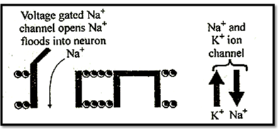
Once the event of depolarization has occurred, a nerve impulse or spike is initiated. Action potential is another name of nerve impulse. This is generated by a change in the sodium ion channels. These channels, and some of the potassium ion channels, are known as voltage gated channel, meaning they can be opened or closed with change in voltage. In resting state these channels are closed due to binding of Ca++.
An action potential is generated by a sudden opening of the sodium gates. Opening of gates increases the permeability of the axon membrane to sodium ions which enter by diffusion.
This increases the number of positive ions inside the axon. A change of -10mV in potential difference from RMP through influx is sufficiently significant to trigger a rapid influx of Na+ ions leading to generation of action potential. This change of -10 mV is called as threshold stimulus.
At the point where membrane (axolemma) is completely depolarised due to rapid influx of Na" ions, the negative potential is first cancelled out and becomes 0 (Depolarisation). This axolemma is called as excited membrane or depolarised membrane. Due to further entry of Na+, the membrane potential "over shoots" beyond the zero and becomes positive upto +30 to +45mV. This "over shoot" peak corresponds to maximum concentration of sodium inside the axon. This potential is called as action potential. In this state, the inner surface of axolemma becomes positively charged and outer' surface becomes negatively charged.
Repolarisation:
After a fraction of second, the sodium gates closed, Depolarisation of the axon membrane causes potassium gates to open, potassium therefore diffuse out of cell:
Since potassium is positively charged, this makes the inside of cell less positive, or more negative and the process of repolarization or return to the original resting potential begins.
The repolarization period returns the cell to its resting potential (-70 mV). The neuron is now prepared to receive another stimulus and conduct it in the same manner.
Sodium pump starts working to maintain the normal resting membrane potential by expelling Na+ and in taking of K+.
The time taken for restoration of resting potential is called refractory period, because during this period the membrane is incapable of receiving another impulse.
Nerve impulse travels as action potential which passes along axon as a wave of depolarization,
The whole process of depolarisation and repolarisation is very fast. It takes only about 1 to 5 milli second (ms).
Open/Operating -,√, Closed -x
Saltatory conduction of nerve impulse:
This type of conduction occur in myelinated fibre.
Myelin is fatty material with a high electrical resistance and act as electrical insulator in the same way as the rubber and plastic covering of electrical wiring.
The combined resistance of the axon membrane and myelin sheath is very high, but where breaks in the myelin sheath occur known as nodes of Ranvier, the resistance to current flow between the axoplasm and the fluid outside the cell is low. It is only at these nodes local circuits are setup.
This means, in effect that the action potential jump from node to node and passes along the myelineted axon faster as compared to the series of small local circuits in a non-myelinated axon. This type of conduction is called saltatory conduction. Leakage of ions takes place only in nodes of Ranvier and less energy is required for saltatory conduction.
Synaptic Transmission
Telodendria of one neuron form synapse with dendron of next neuron.
It is the junctional region between two neurons where information is transferred from one neuron, to another neuron but no protoplasmic connection. Synapse = Pre synaptic knob + synaptic cleft + post synaptic membrane
Telodendria membrane is called pre synaptic membrane & membrane of dendron of other neuron is called as post synaptic membrane. Space between pre and post synaptic membranes is called synaptic deft.
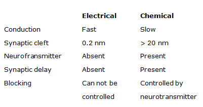
When the AP develop in pre synaptic membrane, it becomes permeable for Ca++ Ca++ enter in pre synaptic membrane & vesicles burst due to the stimulation of Ca++ and release of neurotransmitters (Ach) in synaptic cleft. Ach reaches the post synaptic membrane via synaptic cleft & bind to receptors. It develops excitatory post synaptic potential (EPSP). EPSP develop due to opening of Na+ gatted channels.
Cholinesterase enzyme is found in the synapse.
This enzyme decomposes the Ach into choline & Acetate.
Neuro inhibitory transmitter (GABA) binds with post synaptic membrane to open the Cr gated channels and hyperpolarization of neuron occurs. Now thepotential is called inhibitory post synaptic potential is (IPSP) & further nerve conduction is blocked.
Physiological properties of nerve fibre are detected by cathode ray oscilloscope:
Axqdcindritic - b/w axon & dendron
Axesematic - b/w axon & cyton
Axoaxonic – b/w axon & axon
Synapse
Name synapse was proposed by -Charles Sherrington
Neuron conducts the impulse in the form of electro chemical wave.
Conduction of nerve impulse is unidirectional.
It follows all or none law. Magnitude of response will always be same irrespective of strength of "stimulris' above threshold stimulus.
Velocity of nerve impulse µ Diameter of neuron,
In mammals, the velocity of nerve impulse is 100 to 130 meter/ sec.
This velocity is affected by physical & chemical factor, such as pressure, cold, heat, chloroform and ether etc
Neuroglia / Glial Cells
These arc supporting cells which form a packing substance around the neurons. These are of three types:
|
Astrocytes |
Oligodendrocytes |
Microgliocytes |
|
Origin: Ectodermal in origin |
Ectodermal in origin |
Mesodermal in origin |
|
Morphology: Large cell |
Smaller |
Small cell |
|
Numerous process |
few process |
With branching |
|
Function:- |
|
|
|
1. Provide repair mechanism and replace the damage tissue. |
Formation & preservation of myelin sheath in CNS. |
Scavenger cells of CNS and phagocytic in nature. |
|
2. It forms blood brain barrier. |
|
|
To read more, Buy study materials of Neural Control and Coordination comprising study notes, revision notes, video lectures, previous year solved questions etc. Also browse for more study materials on Biology here.
View courses by askIITians


Design classes One-on-One in your own way with Top IITians/Medical Professionals
Click Here Know More

Complete Self Study Package designed by Industry Leading Experts
Click Here Know More

Live 1-1 coding classes to unleash the Creator in your Child
Click Here Know More


