Nervous System
Table of Content |
|
|

System which regulate the various activities of the body through nerve-impulses is called the nervous system. Through this system the messages are transmitted at a faster rate.
The nervous-system controls and also co-ordinates the various activities of the organs of the animals.
Whole nervous-system of human being is derived from embryonic ectoderm.
Central Nervous System
It includes the brain and the spinal-cord. These are formed from the neural-tube which develops from the ectoderm after the gastrula stage of embryo.
Development of CNS: It develops from neural tube in intrauterine life (I.U.L.). Anterior part of neural tube develops into brain while caudal part of neural tube develops into spinal cord. Brain's approximaieiy 70-80% part of brain develops in 2 year of age & complete development is achieved in 6 years of age & spinal cord develops completely in 4 to 5 years of age.
BRAIN
It is situated in cranial box which is made up of 1 frontal bone, 2 parietal bone, 2 temporal bone, 1 occipital bone. The weight of brain of an adult man is 1400 gm and of female is 1250 gm.
Brain Meninges
Brain is covered by three membranes of connective tissue termed as meninges or menix.
1. Duramater: This is the first and the outermost membrane which is thick, very strong and nonelastic. It is made up of collagen fibres. This membrane is attached with the innermost surface of the cranium.
It is double layer-outer endosteal layer which is closely attached with inner most surface of cranium & no space is found between skull & duramater (No Epidural space). Inner meningeal layer which is related with other meninges of brain, both are vascular. Generally both layers are fused with each other, but at some places these are separated from one another & form a sinus' called cranial venous sinus. These sinuses are filled with venous blood.
2. Arachnoid: It is middle, thin and delicate membrane, made up of connective tissue. It is found only in mammals. It is non vascular layer. In front of cranial venous sinus, it becomes folded, these folds called Arachnoid villi. These villi reabsorb the cerebrospinal fluid (CSF) from sub arachnoid space & pour it into cranial venous sinuses.
3. Piamater: It is inner most, thin and transparent membrane, made up of connective tissue.
Dense network of blood capillaries are found in it, so it is highly vascular. It is firmly adhere to the brain. Piamater & arachnoid layer at some places fuse together to form leptomeninges. Piamater merges into sulci of brain & densely adhere to it. At some places it directly merges in the brain and called telachoroidea Tela choroidea form the choroid plexus' in the ventricles of brain.
Sub Dural Space: Space between duramater & arachnoid. It is filled with serous fluid.
Sub Arachnoid Space: Space between arachnoid & piamater is filled with C.S.F. Cranial nerves also pass through this space. Meningitis: Any inflammation of menix is called as meningitis. It may be caused by viruses bacteria or protozoa.
Cerebrospinal-Fluid (CSF)
This fluid is dear and alkaline in nature just like lymph. It has protein (Albumin, globulin), glucose, cholesterol, urea, bicarbonates, sulphates and chlorides of Na, K. Protein & cholesterol concentration is lesser than plasma & Cl- conc is higher than plasma.
In a healthy man, in 24 hrs, 500 ml of C.S.F is formed & absorbed by arachnoid villi. At a time -total volume of C.S.F. is 150 ml.
CSF is present in ventricle of brain, subarachnoid space of brain & spinal cord.
Formation: Mainly in choroid plexus of lateral ventricles, minor quantity is formed in m= ventricle & IVth ventricle.
Collection of CSF for any investigation is done by lumbar puncture (LP). It is done at L3-L4 region. Spinal anaesthesia is also given by L.P.
Functions of C.S.F.:
Protection of Brain: It act as shock absorbing medium and work as cushion.
It provides buoyancy to the brain, so net weight of the brain is reduced from about 1.4 kg to about 0.18 kg.
Excretion of waste products.
Endocrine medium for the brain to transport hormones to different areas of the brain.
Human brain is divided into three parts –
(1) Fore brain: Cerebrum, Diencephalon.
(2) Mid brain: Consist of many groups of nerve cells called "Nuclei".
(3) Hind Brain: Pons, Cerebellum, Medulla During embryonic stage, brain develops from three hollow vesicles
Cerebrum
It is first & most developed part of brain.
It makes 2/3 part of total brain
Cerebrum consist of two cerebral hemispheres on the dorsal surface. A longitudinal groove is present between two cerebral hemispheres called as median fissure. Both the cerebral hemispheres partially connected with each other by curved thick, nerve fibres is called corpus callosum.
Each cerebral hemisphere is divided into 4 lobes - Anterior, middle, posterior and lateral.
Anterior lobe is also called frontal lobe (largest lobe). Middle lobe is also called parietal lobe. Central sulcus separates frontal lobe from parietal lobe. Lateral lobe or temporal lobe is separated from frontal lobe & parietal lobe by incomplete sulcus called lateral sulcus.
Posterior lobe is also called occipital lobe, it is separated from' parietal lobe, by a sulcus called parieto occipital sulcus.
In right handed person, left hemisphere is dominant while in left handed person right hemisphere is dominant.
Many ridges & grooves are found on dorsal surface of cerebral hemisphere. Ridges are known as gyri while grooves are called sulci. These cover the 2/3 part of cerebrum.
Gyri & sulci are more developed in human being so human being are most intelligent living beings.
Corpus Callosum
It' is largest commissure of brain.
Exclusive feature of mammals. Curved thick-band of white nerve situated between two cerebral hemisphere in the median fissure. Anterior truncated part of corpus callosum is called genu while posterior truncated part is called splenium. A oblique band is formed body of corpus callosum & it goes towards Genu called fornix.
A small cavity is developed among body of callosum. Genu & fornix called' as Vth ventricle or pseudocoel. This ventricle
Is covered by a thin membrane called as septum lucidum.
Diencephalon
It is small and posterior part of fore brain. It is covered by cerebrum. It consists of thalamus, hypothalamus, epithalamus & metathalamus.
(i) Thalamus: It forms upper lateral walls of diencephalon. It forms 80% part of diencephalon . It acts as a relay centre. It receives all sensory impulses from all part of body & these impulses are send to the cerebral cortex.
(ii) Hypothalamus: It forms lower lateral wall of diencephalon.
A cross like structure is found on anterior surface of hypothalamus called as optic chiasma. Pituitary body is attached to middle part of hypothalamus by infundibulum.
Corpus mammillare or Corpus albicans is found on the posterior part of hypothalamus. It is a character of mammalian brain.
Epithalamus: It forms the roof of diencephalon. Pineal body is found in epithalamus.
Metathalamus: It consists of medial geniculate body & lateral geniculate body. It is located in floor of Diencephalon.
Mid brain: It is small & contracted part of brain. Anterior part of mid brain contains two longituidnal myelinated nerve fibres peduncles called cerebral peduncles or crus cercbri or crura cerebri. On the posterior part of mid brain are found four spherical projection called colliculus or optic lobes. Four colliculus are collectively called as corpora quadrigemina (2 upper & 2 lower).
Only 2 colliculus or optic lobes are found in mid brain of frog called as corpora bigemina.
Hind Brain:
3 Parts - (1) Pons (2) Cerebellum
(3) Medulla Oblongata (M.O.)
1. Pons or Pons varolii:
It is small spherical projection, which is situated below the midbrain & upper side of the M.O. It consists of many transverse & longitudinal nerve fibres. Transverse nerve fibres connect with cerebellum.
Longitudinal fibres connect cerebrum to M.O.
2. Cerebellum: Made up of 3 lobe [2 lateral lobe & 1 vermis (divide in 9 segments)].
Both lateral lobes become enlarged & spherical in shape, so lateral lobe of cerebellum are also called as cerebellar hemisphere. Due to this reason, regulation & coordination of voluntary muscle is more developed as compared to other animals.
In terminal part of vermis, one pair of flocculonodular lobes are found. These continue in the form of flocculus. Three cerebellar peduncle are formed, superior cerebellar peduncle attach with mid brain. Middle cerebellar peduncle attach with pons and inferior cerebellar peduncle attaches with M.O.
Medulla Oblongata (M.O.): Posterior part of brain is tubular & cylindrical in shape.
Mid brain, pons & medulla are situated in one axis and called as brain stem.
Internal Structure of Brain
One pair of olfactory lobes are small spherical & solid in human brain. No ventricle is found in it. Both olfactory lobe are separate with each other & are embedded into ventral surface of the both frontal lobe of cerebral hemisphere. Olfactory centre is situated in temporal lobe.
Except midbrain, cerebellum, pons & olfactory lobe complete brain is internally: hollow. Its cavity is lined by ependymal epithelium. (Ciliated Columnar Epithelium).
Cavities of brain are known as ventricles, filled with cerebrospinal fluid (C.S.F.).
In Rabbit, cavity of olfactory lobe is hollow called as 1st ventricle or Rhinocoel. Both Rhinocoel continue in cavity of cerbral hemisphere, known as 2nd ventricle or paracoel or lateral ventricle.
On the posterior side, both paracoel combine with each other & open into cavity of diencephalon through an aperture known as Foramen of Monro.
Cavity of diencephalon is known as 3rd ventricle or Diocoel.
A tent shape space or cavity is present between anterior pons, medulla & posterior cerebellum called IVth ventricle
3rd and 4th ventricle are connected with each other through a hollow tube known as Iter or Aqueduct of sylvius. IVth ventricle continues in the metacoel and metacoel continues in the cavity of spinal cord called neurocoel or central canal.
One aperture is found on dorsal surface of metacoel known as foramen of Magendie.
Two apertures are found on lateral sides of metacoel know as Foramen of Luschka. [1-1]
The CSF of brain comes out from the foramen of Magendie & Luschka & is poured into subarachnoid space.
On dorsal surface of cerebral hemisphere, gray matter becomes more thick, this thick layer of gray matter is known as Cerebral cortex/Neopallium/ Pallium.
Outer part of cerebellum is made up of gray matter while inner part is of white matter. White matter projects outside & forms a branched tree like structure known as Arbor Vitae
Choroid Plexus
Tela choroidea (Piamater which is merged in ventricle)
+ Blood capillaries + Ependymal epithelium
Site: Two major plexuses in lateral ventricles.
2 minor plexuses in m= ventricle
1 minor plexus in IVth ventricle
Function: Formation of CSP by secretion of plasma Some lime (Congenitally or Infection) Iter or aqueduct become blocked therefore improper circulation of CSF or blockage of CSF circulation occur and Intra cranial pressure increases, head becomes enlarged, this condition called Hydrocephalus
Circulation: Prom the ventricles CSF comes into subarachnoid space through foramen of Magendie & foramen of Luschka.
In sub arachnoid space, CSF is absorbed by arachnoid villi which pour it into cranial venous sinus. From venous sinus CSF enters in blood circulation.
Basal nuclei:
Situated in the wall of cerebral hemisphere. Corpus striatum (Caudate nucleus + lentiform nucleus)
+ Amygdaloid body + Claustrum.
Function:
(1) It maintains muscle tone.
(2) It regulates automatic associated movement like swinging of arms during walking
(3) In lower animals, when cerebral cortex is not developed basal nuclei acts as motor centre. Lesion in basal nuclei leads to a disease called as parkinsonism (Rigidity, Tremor at rest).
Reticular activating System:
It is special sensory fibre which is situated in brain stem & further go into Thalamus. It is related to consiousness, alertness & awakening. Therefore it is also called gate keeper of consiousness.
Limbic System: - It is visible like a wish bone, tuning fork or lip like.
Limbic lobe (area of temporal Lobe)
Hippocampus + Hypothalamus including septum + Part of Thalamus + Amygdaloid complex
Functions of Limbic System:
(1) Behaviour, emotion, rage and anger (hypothalamus, amygdaloid body)
(2) Recent memory & short term memory converts into long term memory (Hippocampal lobe)
(3) Food habit (Hypothalamus)
(4) Sexual Behaviour (Hypothalamus)
(5) Olfadion (Hippocampal lobe and Limbic lobe)
Some Analysis Centre Found in the Cerebral Hemisphere
Functions of Brain
1. Olfactory lobe: It is supposed to be centre of smelling power. Its size is small in mammals comparatively because most of its parts become a part of cerebrum. Some animals like sharks and dogs have well developed olfactory lobes.
2. Cerebral hemispheres: It is the most developed part in mammals. It is the most important part of brain because it controls and regulates different parts of brain. This is the centre of conscious senses, will power, voluntary movements, knowledge, memory, speech and thinking, reasoning etc.
Different sense organs send impulses here, and in this part of brain analysis and coordination of impulses is done. Then messages are transferred according to the reactions through voluntary muscles. All the voluntary actions are controlled by cerebral hemispheres.
3. Diencephalon: Its dorsal side is called epithalamus in which pineal body is situated, that controls the sexual maturity of animal.
Thalamus: Act as relay centre for sensory stimulation. In lower animal, cerebral cortex is not developed. Then thalamus act as sensory centre. It is related with RAS.
It is also act as limbic part
Functions of Hypothalamus –
(1) Thermoregulation
(2) Behaviour and emotion
(3) Endocrine control
(4) Biological clock system
(5) ANS control.
These are found centres of animal feelings. Emotions like sleep, anger, intercourse, hate, love, affection, and temperature pain, hunger, thirst and satisfaction are controlled in the hypothalamus.
The regulatory hormones of hypothalamus control the activity of endocrine glands. Modern scientists suppose that hypothalamus is the "master gland" not the pituitary.
Optic chiasma found in the hypothalamus carry optic impulses received from eyes to the cerebral hemispheres, Animal becomes blind if this part is destroyed by chance.
Metathalamus: It is related with MGB & LGB. MGB related with hearing & LGB related with vision. Nerve fibre of concerning place go through Metathalamus.
4. Mid - Brain: Four optic lobes or colliculus present, superior optic lobes are the main centres of pupillary light reflexes. Inferior optic lobes are related acoustic (sound) reflex action.
Crura ccrebri controls the muscles of limbs.
5. Cerebellum: By this portion of hind brain, impulses are received from different voluntary muscles and joints and then controlling of the their movements and their regulation and coordination accordingly is the main function of this part of brain i.e. cerebellum maintains the body balance of persons which take alcohol in excess, their cerebellum gets affected, as a result of that they can not maintain their balance and their walking is disturbed. Thus it is related with fine & skillful voluntary movement & also related with body balance, equillibrium, posture & tone
6. Pons: It regulates the breathing reaction through pneumotaxic centre.
7. Medulla Oblongata: It is the most important part of brain which controls all the involuntary activities of the body. E.g. heart beats, respiration, metabolism, secretory actions of different cells rate of engulfing food etc.
Except this it acts as conduction path for all impulses between spinal cord and-remaining portions of brain. It is also concerned 'with Reflex- Sneezing reflex, salivation reflex, coughing reflex, swallowing reflex, vomiting reflex; yawning reflex.
Spinal Cord
Medulla oblongata comes out from foramen of magnum & continues in neural canal of vertebral column. The continued part of MO is known as Spinal cord. It extends from base of skull to lower vertebra of lumbar. (L1)
Its upper part is wide while lower most part is narrow known as conus-medullaris.
Conus medullaris is present upto L1 vertebra. Terminal part of conus medullaris extend in the form of thread like structure made up-of fibrous connective tissue called filum terminale.
Filum terminale is non-nervous part. Metacoel also continues in spinal cord where it is known as neurocoel or central canal.
Spinal cord is also covered by duramater, arachnoid & piamater. A narrow space is found between vertebra & dura mater known as epidural space;
Length of spinal cord is 45 cm.
Length of filum terminale is 20 cm.
Weight of spinal cord is approximately 35 gm
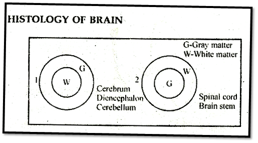
The outer-part of spinal cord is of white matter while inner-part contain gray matter.
On the dorso-lateral& ventro-lateral surface of spinal cord, the gray matter projects outside & forms the one pair dorsal & ventral horn.
Due to formation of dorsal & ventral horn, white matter is divided in 4 segments & segment is known as Funiculus or white column.
Dorsal & ventral horn continue in a tube like (bundle of nerve fibres) structure known as root of dorsal & ventral Horn. In root of dorsal horn, ganglia is present called dorsal root ganglia.
Both root are combined with each other at the place of Intervertebral foramen.
Sensory neurons are found in the dorsal root ganglia which is pseudounipolar in nature & near to intervertebral foramen. Its axon extend & gets embedded into gray matter of spinal cord & sensory nerve fibre come from ganglia & make synapse with ventral root neuron.
Motor neurons are found in the ventral root. Cyton is found in ventral horn while its dendrons are embedded into gray matter of spinal cord where they make synapse with axon of sensory neuron. Axon of motor neuron extends upto intervertebral foramen.
Both sensory & motor nerve fibers combindly come out from intervertebral foramen & form spinal nerve. In some part of spinal cord on both side lateral horns are also found. Also lateral horn cell are found in these horn. From here, nerve fibre come through ventral root & further come into interverbral foramen. These fibre called Ramus communicans.
The group of spinal nerve at the terminal enc (L1) of Spinal cord form tail like structure called cauda equina (horse tail).
Ramus communicans forms ANS.
Spinal nerve & its branches are mixed type except.
Ramus communicans.
(1) It acts as bridge between brain '& organs of the body.
(2) It also provides relay path for the impulses coming from brain.
(3) Spinal cord regulates and conducts the reflex action.
Reflex Action
"Marshal Hall" first observed the reflex actions.
Reflex actions are spontaneous, automatic, involuntay, mechanical responses produced by specific stimulating receptors.
Reflex actions are involuntary actions. Reflex actions are completed very quickly as compared to normal actions. No adverse effect.
It is form of animal behaviour in which The stimulation of a sensory organ (receptor) result in the activity of some organ without the intervention of will.
Reflex actions are of 2 types:
(A) Cranial reflex: These actions are completed by brain. No urgency is required for these actions these are slow actions e.g. watering of mouth to see good food.
(B) Spinal reflex: These actions are completed by spinal cord. Urgency is required for these actions.
These are very fast actions. e.g. Displacement of the leg at the time of pinching by any needle. Classification of reflex actions on the basis of previous experiences:
(A) Conditioned reflex: Previous experience is required to complete these actions e.g. swimming, cycling, dancing, singing etc. These actions were studied first by Evan Pavlov on dog.
Initially these actions are voluntary at the time of learning and after perfection these become involuntary.
(B) Unconditioned reflex: These actions do not require previous experience e.g. sneezing, coughing, yawning, sexual behaviour for opposite sex partner, migration in birds etc.
Reflex Arch
The path of completion of reflex action is called reflex arch.
Sensory fibres carry sensory impulses in the gray matter. These sensory impulses are converted now into motor impulses and reach up to muscles. These muscles show reflex actions for motor impulses obtained from motor neurons, Reflex arch is of two types.
(1) Monosynaptic: In this type of reflex arch, there occurs direct synapse (relation) between sensory and motor neurons. Thus nerve impulse \rave\s through only one synapse ego Stretch reflex
(2) Polysynaptic: In this type of reflex arch, there are found one or more small neurons in between the sensory and motor neurons. These small neurons are called connector neuron or inter neurons or internuncial neurons e.g. withdrawal reflex.
Nerve impulse will have to travel through more than one synapses in this reflex arch.
With drawal Reflex: Sensory neuron supplies the sensation through dorsal root ganglia. Terminal branches of Axon divide in the gray matter & one is supplied by agonist muscle & other is supplied by antagonist muscle. EPSP develop in synapse between motor fibre of agonist muscle & sensory fibre & due to interconnection of interneuron with antagonist muscle neuron, H)SP develop in synapse between colateral branch at sensory fibre and antagonist muscle. Therefore, contraction of agonist muscle and relaxation of the fibre of antagonist muscle.
|
Knee jerk reflex |
Withdrawal reflex |
|
1. No involvement of interneuron. |
Role of interneuron is important. |
|
2. It is an example of monosynaptic reflex. |
Peripheral Nervous System
All the nerves arising from brain and spinal cord are included in peripheral nervous system.
Nerves arising from brain are called cranial nerves and nerves coming out of spinal cord are called spinal nerves.
12-pairs of cranial nerves are found in reptiles, birds and mammals but amphibians and fishes have only 10 pairs of cranial nerves.
In human, I, II and VIII are cranial nerves out of 12 pairs of total, cranial nerves are pure sensory in nature.
III, IV, VI, XI and XII cranial nerves are motor nerves and V, VII, IX & X cranial nerves are mixed type of nerve.
Fibres of autonomous nervous system are found in III, VII, IX & X cranial nerves.
Longest cranial nerve is Vagus nerve.
Largest cranial nerve is Trigeminal nerve.
Smallest cranial nerve is Abducens cranial nerve.
Table: Summary of Human Cranial Nerves:
|
No. |
Name |
Origin |
Distribution |
Nature |
Function |
|
I. |
Olfactory |
Olfactory epithelium |
Enters Olfactory lobe, Extends to temporal lobe. |
Sensory |
Smell |
|
II. |
Optic |
Retina |
Leads to occipital lobe. |
Sensory |
Sight |
|
III. |
Oculomotor |
Midbrain |
Four eye muscles |
Motor |
Movement of eyeball |
|
IV. |
Trochlear (Pathetic) |
Midbrain |
Superior oblique eye muscle. |
Motor |
Movement of eyeball. |
|
V. |
Trigeminal (Dentist nerve) (i) Ophthalmic (ii) Maxillary (iii) Mandibular |
Pons |
Skin of nose, upper eyelids, forehead, scalp, conjunctiva, lacrhymal gland. Mucous membrane of cheeks and upper lip and lower eyelid Lower jaw, lower lip, pinna. |
Mixed -Sensory -Sensory Mixed |
Sensory supply to concerning part - Muscle of Mastication |
|
VI. |
Abducens |
Pons |
lateral rectus eye muscle |
Motor |
Movement of eyeball |
|
VII. |
Facial |
Pons |
Face, neck, taste buds, salivary gland |
Mixed |
Taste (antr 2/3 part of Tongue) Facial expression, saliva secretion. |
|
VIII. |
Auditory (i) Cochlear. (ii) Vestibular |
Pons |
Internal ear |
Sensory |
Hearing and equilibrium. |
|
IX. |
Glossopharyngeal |
Medulla |
Muscles and mucous membrane of pharynx and tongue. |
Mixed |
Taste (postr 1/3 part of tongue) & saliva secretion |
|
X. |
Vagus (Pneumogastric) |
Medulla |
Larynx, lungs, heart, stomach, -intestine |
Mixed |
Visceral sensations and movements. |
|
Xl. |
Accessory spinal |
Medulla |
Motor |
Movement of pharynx, larynx. |
|
|
XII |
Hypoglossal |
Medulla |
Muscles of tongue |
Motor |
Movement of tongue |
Spinal Nerves
In rabbit there are 37 pairs of spinal nerves, while in frog there are 9 or 10 pairs of spinal- nerves.
In Human only 31 pairs of spinal- nerves are found. Each spinal nerve is mixed type and arises from the roots of the horns of gray matter of the spinal cord.
In dorsal root only afferent or sensory fibres and in ventral root efferent or motor fibres are found.
Both the roots after moving for distance in the spinal cord of vertebrates combine with each other and come out from the Inter verterbal foramen in the form of spinal nerves.
As soon as the spinal nerves come out of the inter vertebral foramen they divide into 3 branches:
Autonomic Nervous System
(1) The autonomic nervous system is that part of the peripheral nervous system which controls activities inside body that are normally involuntary, such as heart beat, peristalsis, sweating etc.
(2 It consists of motor neuron passing to the smooth muscle of internal organs. Smooth muscles are involuntary muscles. Most of the activities of the autonomic nervous system is controlled within the spinal cord or brain by reflexes known as visceral reflexes and does not involve the conscious control of higher centres of the brain.
Overall control of the autonomic nervous system is maintained, however, by centres in the medulla (a part of the hind brain) and hypothalamus.
These receive and integrate sensory information and coordinate this with information from other parts of the nervous system to produce the appropriate response.
ANS plays an important role in maintaining the constant internal environment (homeostasis).
The autonomic nervous system is composed of two type of neurons, a preganglionic neuron (myelinated which leaves the central nervous system in the venteral root before synapsing several postganglionic neurons (non myelinated) leading to effector (concermng organs)
Mechanism Sites of ANS
Involuntary muscles, exocrine glands. Blood vessels, skin (Pilomotor muscles) Blood vessels, Sweat glands
There are the two division of the autonomic nervous system- the sympathetic (SNS) and the parasympathetic (PNS).
(i) Sympathetic system is related with such visceral reactions. which increases the protection of body adverse atmospheric conditions along with calorie consumption.
(ii) Parasympathetic system is related with those reactions in which energy is conserved.
In this way, autonomic nervous system controls the activites of visceral organs double sided i.e. antagonist to each other.
|
|
Sympathetic NS |
|
Parasympathetic NS |
|
1 |
Thoracico lumbar outflow (T1 to L3) (Ramus communicans of T1 to L3) |
1 |
Cranio sacral outflow (cranial nerves) 3, 7, 9, 10 and sacral's ramus communicans 2, 3, 4 |
|
2 |
Just lateral to vertebral column sympathetic trunks are there on both sides· (each made up of 22 ganglia) |
2 |
Ganglia are situated separately either near the organ or surface of organ. |
|
3 |
Preganglionic nerve fibres (Ramus communicans of spinal nerves) are smaller than post ganglionic nerve fibres (arises from sympathetic trunk or ganglia to organs) |
3 |
Preganglionic nerve fibres are longer than postganglionic nerve fibres. |
|
4 |
Preganglionic nerve fibres are cholinergic (filled with acetylcholine). and post ganglionic nerve, fibres are adrenergic (filled with noradrenaline) except sweat gland which have cholinergic postganglionic nerve fibres. |
4 |
Both pre and post ganglionic nerve fiber are cholinergic |
|
5 |
Supply (a) Cervical ganglia, head, face (b) Cervical and thoracic ganglia heart (c) Thoracic ganglia ® Trachea, bronchi and lungs (d) Preganglionic nerve fibres of abdominal part do not form synapse in sympathetic trunk. They form synapse in prevertebral ganglia (coeliac, superior mesenteric and inferior mesenteric). Coeliac and superior mesenteric supply Stomach to proximal colon Inferior mesenteric supply - distal colon, urinogenital organ, rectum and anal canal. Exception: Adrenal medulla receive preganglionic nerve directly. (No any postganglionic fibres) |
5 |
Supply (a) Cranial nerve - iii- iris, ciliary muscles vii- Tear glands, palate, pharynx, submandibular and sublingual glands: ix- Parotid gland, buccal cavity x- Neck to proximal colon. (b) Sacral 2, 3,4 distal colon, rectum, anal canal and urinogenitall organ
|
|
6 |
Preganglionic nerve fibres are made up of white ramus communicans and postganglionic nerve fibres are made up of gray ramus communicans. |
6 |
|
- Nervous Control of Visceral Organs
|
Name of Visceral Organs |
Affect of sympathetic nervous system |
Affect of parasympathetic nervous system |
|
1. Secretion |
Acetyl choline + sympathetin |
Only acetylcholine |
|
2. Iris of eye |
Dilates pupils |
Constricts pupils |
|
3. Tear glands or lachrymal glands |
Stimulates secretion of lachrymal glands |
Inhibits secretion of lachrymal glands |
|
4. Heart |
Increases the rate of cardiac contraction i.e. accelerates heart beat |
Inhibits the rate of cardiac contraction i.e. retards heart beat. |
|
5. Secretion of adrenal gland |
Stimulates adrenal secretion |
Inhibits adrenal secretion. |
|
6. Salivary secretion |
Inhibits the secretion of salivary and digestive glands |
Stimulates the secretion of salivary and digestive glands |
|
7. Blood vessels |
Constricts cutaneous blood vessels, which causes increased blood pressure but dilates blood vessels of brain, lungs, heart, striated muscles. Increases RBC count in blood. |
Dilates all blood vessels (except coronary vessels) decreasing blood pressure |
|
8. Lungs, trachea and bronchi |
Dilates trachea, bronchi & lungs for easy breathing |
Constricts these organs during normal breathing. |
|
9. Alimentary canal |
Inhibits peristalsis of alimentary canal. |
Stimulates the peristalsis of alimentary canal |
|
10. Digestive glands. |
Inhibits the secretion of these glands |
Stimulates the secretion of the glands |
|
11. Sweat glands |
Stimulates secretion of sweat. |
Inhibits secretion of sweat. |
|
12. Arrector pilli muscles |
Stimulates contraction of these muscles of skin, causing goose flesh |
Relaxes arrector pilli muscles. |
|
13. Urinary bladder |
Relaxes the muscles of urinary bladder. |
Contracts the muscles for ejaculation of urine (Micturition). |
|
14. Anal sphincter |
Relaxes anal sphincter and opens the anus (Deraecation). |
|
|
15. External genitalia |
Ejaculation , |
Erection |
|
16. Basal metabolic rate |
Accelerates BMR. |
Retards BMR. |
Comparative Account of Nervous System in Rabbit and Human
EYE
Our paired eyes are located in sockets of the skull called orbits.
The adult human eye ball is nearly a spherical structure. The wall of the eye ball is composed of three layers. The external layer is composed of a dense connective tissue and is called the sclera. The anterior portion of this layer is called the cornea. The middle layer, choroid, contains many blood vessels and looks bluish in colour. The choroid layer is thin over the posterior two-thirds of the eye ball, but it becomes thick in the anterior part to form the ciliary body. The ciliary body itself continues forward to form a pigmented and opaque structure called the iris which is the visible coloured portion of the eye.
The eye ball contains a transparent crystalline lens which is held in place by ligaments attached to the ciliary body. In front of the lens, the aperture surrounded by the iris is called the pupil. The diameter of the pupil is regulated by the muscle fibres of iris. The inner layer is the 'retina and it contains three layers of cells - from inside to outside - ganglion cells, bipolar cells and photoreceptor cells.
There are two types of photoreceptor cells, namely, rods and cones. These cells contain the light sensitive proteins called the photopigments. The daylight (photopic) vision and colour vision are functions of cones and the twilight (scotopic) vision is the function of the rods.
The rods contain a purplish-red protein called the rhodopsin or visual purple, which contains a derivative of Vitamin A.
In the human eye, there are three types of cones which possess their own characteristic photopigments that respond to red, green and blue lights. The sensations of different colours are produced by various combinations of these cones and their photopigments. When these cones are stimulated equally, a sensation of white light is produced.
The optic nerves leave the eye and the retinal blood vessels enter it at, a point medial to and slightly above the posterior pole of the eye ball. Photoreceptor cells are not present in that region and hence it is called the blind spot. At the posterior pole of the eye lateral to the' blind spot, there is a yellowish pigmented spot called macula lutea with a central pit called the fovea. The fovea is a thinnedout portion of the retina where only the cones are densely packed. It is the point where the visual acuity (resolution) is the greatest.
The space between the cornea and the lens is called the aqueous chamber and contains a thin watery fluid called aqueous humor. The space between the lens and the' retina is called the vitreous chamber and is filled with a transparent gel called vitreous humor.
Largest eye ball is found in the deer.
Smallest eye ball is found in monkey.
Size of eye ball in human beings. New born = 1.75 cm, Adult = 2-2.5 cm
Sharpest vision found in the Eagle (In day time) & in night time - Owl.
Lowest sight - Monkey. [Minimum focal length]. Vision of human being - Binocular stereoscopic (3 dimensional vision).
In most of the animals monocular vision is found. e.g. Rat, Horse, Rabbit.
Minimum distance for proper vision of eye = 25 cm, Maximum sensitivity of eye is for 5000 A.
Telescopic eye is found in birds. (large focal length.)
Colour vision is found in Humans, Monkey, Lizard, Snakes, Birds, Fresh water fishes.
In Hen, only cones are found. In Owl, only rods are found.
In human beings, both rods and cones are found [Rods (120 million) and Cones (7 million)]
Black and white vision due to rods & colour vision is due to cones
Dim light vision (Black & white vision) is due to rods & colour vision due to cones.
Eye ball is protected by –
Eyebrows
Eye lids & Eye lashes
Glands
Three eye lid present in the rabbit.
First -upper eye lid
Third -Nictitating membrane.
Third eye lid is vestigeal in human being known as plica semilunaris. In frog and other amphibiar sclerotic layer of eye ball is cartilaginous. Eye lids are absent in snake. Immovable eye lids are present in the fishes. Cornea and lens has no blood and no nerves supply. Image on retina is inverted & real
Refractive Error of Eye
1. Myopia: Image form infront of retina and far objects are not clearly seen.
Reason - The increase curvature of cornea or increase convexity' of lens.
Prevention - By biconcave lens corrected the myopia.
2.Hypermetropia: Image is formed behind the retina. So near objects are not seen clearly. Reason -In this, lens become flat or decrease curvature of cornea decrease convexity of lens. Eye of new born baby is "hypermetropic.
Prevention - It is corrected by convex lens.
3. Presbyopla: After 35 to 40 year elasticity of lens decrease so hypermetropia occur. Hypermetropia due to ageing is called presbyopia. Correctted by convex lens.
4. Astigmatism: Due to different curvature of lens at the different place.
Overall image is not form at the yellow spot. Corrected by cylindrical lens. Due to dislocation of lens or trauma.
Other disease:
1. Cataract: After 60 years lens become opaque due to destruction of cysteins and Glutathione amino acids. In India most common cause of blindness. (80%)
Treatment-Removal of cataractal lens and replace by intraocular' lens.
2. Glaucoma: At the junction of cornea and sclera there is a canal called schlemn canal. This canal drains out aqueous humour into the veins. Aqueous humour formed by the 'blood vessels of ciliary process. Some time Schlemm canal block drainage of aqueous humour does not occur. Hence, aqueous humour gets collected in anterior and posterior chamber of eye. So 101 (Intraocular pressure) increases (normally 1023 mm of Hg) and condition is called Glaucoma.
3. Trachoma: Caused by chlamydia- also called LPT virus or Basophilic virus. In this inflammation of conjunctive occurs called conjuctivitis. Eye become red called swimming pool disease. Eye-flu due to influenza virus.
4. Hystagmus: Pendular movement of eye ball occurs.
The Ear
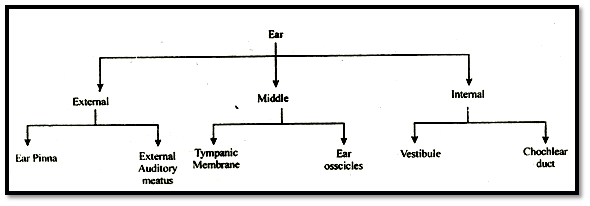
The outer ear consists of the pinna and external auditory meatus (canal). The pinna collects the vibrations in the air which produce sound. The external auditory meatus leads inwards and extends up to the tympanic membrane (the ear drum). There are very fine hairs and wax secreting sebaceous glands in the skin of the pinna and the meatus. The tympanic membrane is composed of connective tissues covered with skin outside and with mucus membrane inside.
The middle ear contains three ossicles called malleus, incus and stapes which are attached to one another in a chain-like -fashion. The malleus is attached to the tympanic membrane and the stapes is attached to the oval window of the cochlea. The ear ossicles increases the efficiency of transmission of sound-waves to the inner ear. An Eustachian tube connects the middle ear cavity with the pharynx. The Eustachian tube helps in equalising the pressures on either sides of the ear drum.
The ears perform two sensory functions, hearing and maintenance of body balance.
The ear can be divided into three major sections called the outer ear, the middle ear and the inner ear basilar, divide the surounding perilymph filled bony labyrinth into an upper scala vestibuli and a lower scala tympani. The space within cochlea called scala media is filled with endolymph. At the base of the cochlea, the scala vestibuli ends at the oval window, while the scala tympani terminates at the round window which opens to the middle ear.
The organ of corti is a structure located on the basilar membrane which contains hair cells that act as auditory receptors. The hair cells are present in rows on the internal side of the organ of corti. The basal end of the hair cell is in close contact with the afferent nerve fibres. A large number of processes called stereo cilia are projected from the apical part of each hair cell. Above the rows of the hair cells is a thin elastic membrane called tectorial membrane.
The inner ear contains a complex system called vestibular apparatus, located above the cochlea. The vestibular apparatus is composed of three semicircular canals and the otolith organ consisting of the saccule and utricle. Each semi-circular canal lies in a different plane at right angles to each other. The membranous canals are suspended in the perilymph of the bony canals. The base of canals is swollen and is called ampulla, which contains a projecting ridge-called crista ampullaris which has half cells. The saccule and utricle contain a projecting ridge called macula. The crista and macula are the specific receptors of the vestibular of paratus responsible for maintenance of balance of the body and posture.
NOTE:
Merrier's syndrome is -disease associated with middle ear:
Bat has thickest auditory nerve made up of 30000 neurons.
The turtle, amyda lacks rods completely.
Umbraculum is the covering as the cornea of camel.
Fresh water fishes have prophyropsin in rods and cyartopsin in the cones.
Man can hear 20-20,000 Cycles per second.
Special Points
In the brain of frog only 2 skull meninges are present. The middle meninge that is the arachnoid is absent.
In rabbit, man and mammals- 3 skull meninges are present.
Increase in the amount of cerebro- spinal fluid is a diseased condition termed as the Hydrocephalus
"Meningitis". It arises due to infection or inflammation on injury in the meninges. Infection may be due to virus or bacteria or both.
Piamater is the most vascular and conducting and provides nutrition.
The power of regeneration is very less in a neuron.
The optic lobes of frog are hollow and in them optocoel cavity is found.
In frog, 2 optic- lobes are present. These are hollow and termed as. Corpora- bigemina. In mammals, 4 solid optic - lobes are present.
Around the brain of fishes, only one menix is found called "Menix- primitiva",
The valve of vieussens joins the optic-lobes with the cerebellum.
Cerebral - cortex is made up of gray - matter and is divided into 3 areas:
Sensory area
Motor area
Associated area
The sensory and associated areas determine the shape. colour, sound, taste and smell of any object.
Motor area regulates muscular - contraction.
Broca's area: It is known as motor speech area.
Broca's area is present in the lateral part of the frontal- lobe of the cerebrum. This area makes aware of language; and translates the written words into speech.
If Broca's area gets destroyed the animal becomes unable to speak.
The temporal- lobes of cerebrum regulates the mechanism of hearing.
Cerebrum is the centre of following:
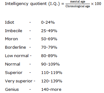
Intelligence
Emotion
Will- power
Memory
Consciousness
Experience
Knowledge
Voluntary control
Laughing and weeping
Defaecation and micturition.
Diencephalon is the centre of carbohydrate - metabolism and fat- metabolism.
In rabbit "Swammerdarn's glands" are absent. These glands are present at the origin plate of spinal nerves is vertebrates & present in frog. It provide extra supply of Ca++ for synaptic transmission.
Cerebellum is made up of three layers and in the middle of cerebellum of brain lobes of flask- shaped cells are found called the "Purkinje-cells".
In the nerve- cells. "centrosome" are absent so once formed in development, the nerve-cells do not divide and remain in inter-phase stage throughout their lifer and grow in size with the growth of the body.
The velocity of nerve- impulse is 5 to 50 times more faster in myelinated nerve-fibres than in non-myelinated nerve-fibres.
In mammals the speed of nerve impulse is 100-130 m/sec (maximum). In frog the speed of nerve impulse is 30 m/sec. In reptiles the speed is 15 to 35 m/sec.
Acetylcholinesterase enzyme helps in the dissociation of Acetylcholine.
In the form of inhibitory neuro- hormons-GABA are present.
Acetylcholine is synthesized by the Mitochondria.
For the conduction of nerve- impulses, Na+ is necessary.
The marking of brain- waves is done through E.E G i.e Electro- encephalo gram.
In embryonal - stages inside the brain the layer of gray- matter is towards inside and that of white - matter is towards outside. In adults, this arrangement continues in the spinal-cord but during the development of the wall of the brain the gray matter is transferred outside.
In 1 minute 750ml of blood is conducted to the human- brain.
Corpus callosum is the largest commissure of the brain.
Largest cranial venous sinus is cavernous venous sinus which is situated in the middle of cranial fossa.
All pre ganglionic sympathetic nerve fibres are myelinated while post ganglionic nerve fibre are non mvelinatete.
In PNS myelinogenesis process is occurcd by neurilemma (Schwan cells) while in CNS myelinogenesis process occured by oligodendrocytes {Neuroglia}
Alzheimer's disease - In this disease, the cerebral cortex is atrophied and ultimately the ventricle enlarge. Symptoms consist loss of memory particularly recent memory. Alzheimer disease is more common in Down's syndrome.
Treatment - No effective treatment.
Stroke - May be caused by haemorrhage into the brain.
Symptoms -Unconsiousncss.
Treatment-Intravenous tissue plasrninogen activator.
Epilepsy - Epilepsy is characterized by short, recurrent periodic attack of motor sensory or psychologic malfunction.
Cause - due to abnormal discharge of cerebral neurons.
Symptoms - seizures, unconsiousness. Involuntary contraction of muscle.
Treatment - Anti epileptic drugs.
In human brain more than 100 billion neuroosilre present.
Each neuron cell connect with 25,000 other cell
Phrenic nerve is branch of cervical plexues which supply diaphragm.
Malathionc: This substance is used as insecticide and it destroy the acetyl cholinesterase in synapse area.
Botulism: It is food poisoning disease and it produces by Clostridium botulinum bacterium. This bacteria release neurotoxin.
Curare is drug which blocks acetylcholine receptor on skeletal muscle, so that it can be used by a surgeon for keeping a muscle relaxed during operation.
Corpus striatum regulates planning and execution of stereotyped movement.
The region between the thalamus and spinal cord is referred to as brain stem.
All impulse traversing a given neuron have the same amplitude, differing only in the frequency of impulse. Glycine is neuro inhibitory hormone present in spinal cord.
Glutamate is excitatory amino acid.
Trigeminal nerve is also called "the dentists nerve" because' the dentists desensitizes this nerve with some anaesthetic before pulling out the troubling tooth.
To read more, Buy study materials of Neural Control and Coordination comprising study notes, revision notes, video lectures, previous year solved questions etc. Also browse for more study materials on Biology here.
View courses by askIITians


Design classes One-on-One in your own way with Top IITians/Medical Professionals
Click Here Know More

Complete Self Study Package designed by Industry Leading Experts
Click Here Know More

Live 1-1 coding classes to unleash the Creator in your Child
Click Here Know More


