Endomembrane System-I
Table of Content |
The Endoplasmic Reticulum (ER)
-
 A network or reticulum of tiny tubular structures scattered in the cytoplasm that is called the endoplasmic reticulum (ER).
 -
Garnier (1897) was first to observe the ergastoplasm in a cell. The ER was first noted by Porter, Claude, and Fullman in 1945 as a network.
-
It was named by Porter in 1953.
-
The ER is present in almost all eukaryotic cells. A few cells such as ova, embryonic cells, and mature RBCs, however, lack ER. It is also absent in prokaryotic cell. In rapidly dividing cells endoplasmic reticulum is poorly developed.
-
The ER is made up' of three components. All the three structures are bound by a single unit membrane.
-
Cisternae: These are flattened, unbranched structures. They lie in stacks (piles) parallel to one another. They bear ribosomes. They contain glycoproteins named ribophorin-Land ribophorin II that bind the ribosomes. Found in protein forming cells.
-
Vesicles: These are oval or rounded, vacuole like elements, scattered in cytoplasm. These are also studded with ribosomes.
-
Tubules: Wider, tubular, branched elements mainly present near the cell membrane. They are free from ribosomes. These are more in lipid forming cells.
-
The ER often shows ribosomes attached to their outer surface. The endoplasmic reticulum bearing ribosomes on their surface is called rough endoplasmic reticulum (RER). In the absence of ribosomes they appear smooth and are called smooth endoplasmic reticulum (SER).
-
RER is frequently observed in the cells actively involved in protein synthesis and secretion. They are extensive and continuous with the outer membrane of the nucleus.
-
The smooth endoplasmic reticulum is the major site for synthesis of lipid. In animal cells lipid-like steroidal hormones are synthesised in SER.
Golgi Apparatus
-
 Camillo Golgi (1898) first observed densely stained reticular structures near the nucleus. These were later named Golgi bodies after him. They consist of many flat, disc-shaped sacs or cisternae of 0.5 mm to 1mm diameter. These are stacked parallel to each other. Varied numbers of cisternae are present in a Golgi complex.
 -
It is present in all eukaryotic cells. In plants, these are scattered irregularly in the cytoplasm and called as “dictyosomes".
-
These are absent in bacteria and blue green algae, RBCs, spermatozoa of bryophytes and pteridophytes, and sieve tube cells of phloem of angiosperm. The number of golgi body increased during cell division.
-
Average number 10-20 per cell. Golgi body surrounded by a zone of protoplasm which is devoid of cell organelles called zone of exclusion (Morre, 1977).
-
Under transmission electron microscope the structure of golgi bodies was-study by Dalton and Felix (1954), golgi body is made of 4 parts.
(1) Cisternae: Golgi apparatus is made up of stack of flat sac like structure called cisternae. The margins of each cisterna are gently curved so that the entire golgi body takes on a cup like appearance. The golgi body has a definite polarity. The-cisternae at the convex end of the dictyosome comprises forming face (F. face) or cis face. While the cisternae at the concave end comprises the maturing face (M. face) or trans face. The forming face is located next to either the nucleus or endoplasmic reticulum. The maturing face is usually directed towards the plasma membranes. IUs the functional unit of golgi body.
(2) Tubules: These arise due to fenestration of cisternae and it forms a complex of network.
(3) Secretory vesicles: These are small sized components each about 40 Å in diameter presents along convex surface of edges of cisternae. These are smooth and coated type of vesicles.
(4) Golgian vacuoles:
-
They are expanded part of the cisternae which have become modified to form vacuoles. The vacuoles 'develop from the concave or maturing face.
-
Oolgian vacuoles contain amorphous or granular substance. Some of the golgian vacuoles function as lysosomes
-
The Golgi cisternae are concentrically arranged near the nucleus with distinct convex cis or the forming face and concave trans or the maturing face.
-
The cis and the trans faces of the organelle are entirely different, but interconnected.
-
The golgi apparatus principally performs the function of packaging materials, to be delivered either -to the intra-cellular targets or secreted outside the cell.
-
Materials to be packaged in the form of vesicles from the ER fuse with the cis face of the golgi apparatus and move towards the maturing face.
-
A number of proteins synthesised by ribosomes on the endoplasmic reticulum are modified in the cisternae of the Golgi apparatus before they are released from its trans face.
-
Golgi apparatus is the important site of formation of glycoproteins and glycolipids.
Lysosomes
-
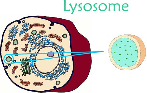
 Lysosomes are electron microscopic, vesicular structures of the cytoplasm, bounded by a single membrane (lipoproteinous) which are involved in intracellular digestive activities, contains hydrolytic enzymes, so called lysosomes.
 -
These were first discovered by a Belgian biochemist, Christian de Duve (1955) in the liver cells and were earlier named pericanalicular dense bodies.
-
Terms Lysosome was given by Novikoff under the study of electron microscope.
-
Matile (1964) was first to demonstrate their presence in plants, particularly in the fungus Neurospora. Polymorphism in lysosomes were described by De Robertis et al (1971).
-
These are absent from the prokaryotes but are present in all eukaryotic animal cells except mammalian RBCs. They have been recorded in fungi, Euglena, cotton and pea seeds.
-
These are' membrane bound vesicular structures formed by the process of packaging in the golgi apparatus. The isolated lysosomal vesicles have been found to be very rich in almost all types of hydrolytic enzymes (hydrolases - lipases, proteases, carbohydrases) optimally active at the acidic pH. These enzymes are capable of digesting carbohydrates, proteins, lipids and nucleic acids.
Types of Lysosomes
On the basis of their contents, four types of lysosomes are recognised.
-
Primary Lysosomes: A newly formed lysosome contains enzymes only. It is called the primary lysosomes. Its enzymes are probably in an inactive state.
-
Secondary Lysosomes: When some material to be digested enters a primary lysosome, the latter is named the secondary lysosome, or phagolysosome or digestive vacuole, or heterophagosome.
-
Tertiary lysosomes/Residual bodies: A secondary lysosome containing indigestible matter is known as the residual bodies or tertiary lysosome. The latter meets the 'cell by exocytosis (ephagy).
-
Autophagosomes/Autolysosomes: A cell may digest its own organelles, such as mitochondria, ER. This process is called autophagy. These are formed of primary lysosomes. The acid hydrolases of lysosomes digest the organelles thus, it is called autophagosome. The lysosome are sometimes called disposal units/suicidal bags. Sometime they get burst and cause the destruction of cell or tissue.
Vacuoles
-
The vacuole is the membrane-bound space found in the cytoplasm.
-
It contains water, sap, excretory product and other materials not useful for the cell.
-
The vacuole is bound by a single membrane called tonoplast. In plant cells the vacuoles can occupy up to 90 per cent of the volume of the cell.
-
In plants, the tonoplast facilitates the transport of a number of ions and other materials against concentration gradients into the vacuole; hence their concentration is significantly higher in the vacuole than in the cytoplasm.
-
In Amoeba the contractile vacuole is important for excretion. In many cells, as in protists, food vacuoles are formed by engulfing the food particles.
Mitochondria
-
Mitochondria (sing: mitochondrion), unless specifically stained, are not easily visible under the microscope.
-
Mitochondria are also called chondriosoine, chondrioplast, plasmosomes, plastosomes and plastochondriane.
-
These were first observed in striated muscles (Voluntary) of insects as granules by Kolliker (1880), he called them sarcosomes.
-
Michaelis (1898) demonstrated that mitochondria play a significant role in respiration.
-
Bensley and Hoerr (1934) isolated mitochondria from liver cells.
-
Seekevitz called them "Power house of the cell"
-
Nass and Afzelius (1965) observed first DNA in mitochondria.
-
Minimum number of mitochondria is one in Microasterias, Trypanosoma, Chlorella, Chlamydomonas (green alga) and Micromonas.
-
Maximum numbers are found (up to 500000) in flight muscle cell, (up to 50000) in giant Amoeba called Chaos - Chaos. These are 25 in human sperm, 300 - 400 in kidney cells and 1000 - 1600 in liver cells.
-
Size of mitochondria: Average size is 0.5–1.00 mm and length up to 1 – 10 mm.
(i) Smallest sized mitochondria in yeast cells 1mm3
(ii) Largest sized are found in oocytes of Rana pipiens and are 20 – 40 mm
(iii) A dye for staining mitochondria is Janus B – green.
Enzymes of Mitochondria
(1) Outer membrane: Monoamine oxidase, glycero phosphatase, acyl transferase, phospholipase A.
(2) Inner membrane: Cytochrome b.c1.c.a, (cyt.b, cyt.c1, cyt.c, cyt.a, cyt.a3 NADH, dehydrogenase, succinate dehydrogenase, ubiquinone, flavoprotein, ATPase.
(3) Perimitochondrial space: Adenylate kinase, nucleoside diphosphokinase,
(4) Inner matrix: Pyruvate dehydrogenase, citrate synthase, aconitase, isocitrate dehydrogenase, fumarase, a-Ketoglutarate dehydrogenase, malate dehydrogenase.
Plastids
-
Definition: Plastids are semiautonomous organelles having DNA, RNA, Ribosomes and double membrane envelope which store or synthesize various types of organic compounds as ATP and NADPH + H+ etc. These are largest cell organelles in plant cell.
-
Haeckel (1865) discovered plastid, but the term was first time used by Schimper (1883).
-
A well organised system of grana and stroma in plastid of normal barley plant was reported by de Von Wettstein.
-
Park and Biggins (1964) gave the concept of quantasomes.
-
The term chlorophyll was given by Pelletier and Caventou, and structural details were given by Willstatter and Stall.
-
Ris and Plaut (1962) reported DNA in chloroplast and was called plastidome.
-
Plastids are found in all plant cells and in euglenoides. These are easily observed under the microscope as they are large.
-
They bear some specific pigments, thus imparting specific colours to the plants. Based on the type of pigments plastids can be classified into chloroplasts, chromoplasts and leucoplasts.
-
The chloroplasts contain chlorophyll and carotenoid pigments which are responsible for trapping light energy essential for photosynthesis.
-
In the chromoplasts fat soluble carotenoid pigments like carotene, xanthophylls and others' are present.
-
This gives the part of the plant a yellow, orange or red colour. The leucoplasts are the colourless plastids of varied shapes and sizes with stored nutrients:
-
Amyloplasts store carbohydrates (starch), e.g., potato; elaioplasts store oils and fats whereas the aleuroplasts store proteins.
Pigments of chloroplast
Chlorophyll a: C55 H72 O5N4 Mg (with methyl group)
Chlorophyll b: Css H70 O6N4 Mg (with aldehyde group)
Chlorophyll c: C35H32 O5 N4 Mg
Chlorophyll d: C54 H70 O6N4 Mg
-
Majority of the chloroplasts of the green plants are found in the mesophyll cells of the leaves.
-
These are lens-shaped, oval, spherical, discoid or even ribbon-like organelles having variable length (5-10mm) and width (2-4mm). Their number varies from 1 per cell of the Chlamydomonas, a green alga to 20-40 per cell in the mesophyll.
-
Like mitochondria, the chloroplasts are also double membrane bound. Of the two, the inner chloroplast membrane is relatively less permeable.
-
The space limited by the inner membrane of the chloroplast is called the stroma.
-
A number of organised flattened membranous sacs called the thylakoids, are present in the stroma. Thylakoids are arranged in stacks like the piles of coins called grana (singular: granum) or the inter grana thylakoids.
-
In addition, there are flat membranous tubules called the stroma lamellae connecting the , thylakoids of the different grana.
-
The membrane of the thylakoids encloses a space called a lumen. The stroma of the chloroplast contains enzymes required for the synthesis of carbohydrates and proteins.
-
It also contains small, double-stranded circular DNA molecules and ribosomes. Chlorophyll pigments are present in the thylakoids.
-
The ribosomes of the chloroplasts are smaller (70S) than the cytoplasmic ribosomes (80S).
Types of Plastids
According to Schimper, Plastids are of 3 types: Leucoplasts, Chromoplasts and Chloroplasts.
Leucoplasts
They are colourless plastids which generally occur near the nucleus in nongreen cells and possess internal lamellae. Grana and photosynthetic pigments are absent. They mainly store food materials and occur in the cells not exposed to sunlight e.g., seeds underground stems, roots, tubers, rhizomes etc. These are of three types.
(i) Amyloplast : Synthesize and store starch grains. e.g., potato tubers, wheat and rice grains.
(ii) Elaioplast (Lipidoplast, Oleoplast) : They store lipids and oils e.g. castor endosperm, tube rose, etc.
(iii) Aleuroplast (Proteinoplast) : Store proteins e.g., aleurone cells of maize grains.
Chromoplasts
-
Coloured plastids other than green are known as chromoplasts.
-
These are present in petals and fruits, imparting different colours (red, yellow, orange etc) for attracting insects and animals. These also carry on photosynthesis.
-
These may arise from the chloroplasts due to replacement of chlorophyll by other pigments e.g. tomato and chillies or from leucoplasts by the development of pigments.
-
All colours (except green) are produced by flavins, flavenoids and cyanin. Cyanin pigment is of two types one is anthocyanin (blue) and another is erythrocyanin (red). Anthocyanin express different colours on different pH value. These are variously coloured e.g. in flowers. They give colour to petals and help in pollination. They are water soluble. They are found in cell sap.
-
Green tomatoes and chillies turn red on ripening because of replacement of chlorophyll molecule in chloroplasts by the red pigment lycopene in tomato and capsanthin in chillies. Thus, chloroplasts are changed into chromatophores.
Chloroplast
Discovered by Sachs and named by Schimper. They are greenish plastids which possess photosynthetic pigments.


Q.1 - A cell homogenate is subjected to ultra centrifugation. What fraction would be separated at 10,000g × 20 minutes
(a) Ribosome and Microsome
(b) Mitochondria and Lysosome
(c) Nucleus and Nucleolus
(d) Endoplasmic reticulum
Q.2 - The order of sedimentation of subcellular structures during differential centrifugation is
(a) Lysosome, mitochondria, ribosome
(b) Mitochondria, nucleus, lysosome
(c) Nucleus, mitochondria, lysosome
(d) Ribosome, nucleus, mitochondria
Q.3 - Organelle not possible to observe without electron microscope is
(a) Chloroplast
(b) Ribosome
(c) Mitochondrion
(d) Nucleolus
Q.4 - Which of the following is absent in prokaryotes
(a) DNA
(b) RNA
(c) Plasma membrane
(d) Mitochondria
Q.5 - Cell organelles found only in plants
(a) Golgi complex
(b) Mitochondria
(c) Plastids
(d) Ribosomes
Q.6 - Which one of the following pairs is correctly matched?
(a) Microsomes - Participate in the process of photosynthesis
(b) Lysosomes - Involved in synthesizing amino acids
(c) Endo. Reticulum - Plays role in the formation of a new nuclear membrane during cell division
(d) Centrosome - Provide enzymes required in the digestive process
Q.7 - All plant cells normally possess
(a) Middle lamella
(b) Primary wall
(c) Lysosomes
(d) Centrioles
Q.8 - Non-living substance in protoplasm is
(a) Carbohydrate
(b) Ribosome
(c) Mitochondria
(d) Plastids


|
Q.1 |
Q.2 |
Q.3 |
Q.4 |
|
b |
c |
b |
|
|
Q.5 |
Q.6 |
Q.7 |
Q.8 |
|
c |
c |
b |
a |
Related Resources
-
Click here to refer the Useful Books of Biology for NEET (AIPMT)
-
Click here for study material on Cell – the unit of life
To read more, Buy study materials of Cell: The Unit of Life comprising study notes, revision notes, video lectures, previous year solved questions etc. Also browse for more study materials on Biology here.
View courses by askIITians


Design classes One-on-One in your own way with Top IITians/Medical Professionals
Click Here Know More

Complete Self Study Package designed by Industry Leading Experts
Click Here Know More

Live 1-1 coding classes to unleash the Creator in your Child
Click Here Know More


