Kingdom Fungi
Table of Content |

 - The science dealing with the study of fungi is called as mycology.
- The knowledge of fungi to mankind dates back to prehistoric times.
- Clausius, 1601 may be regarded as one of the earliest writers to describe fungi.
- Bauhin (1623) also included the account of known fungal forms in his book Pinax Theatric Botanica.
- The fast systematic account of fungi came from Pier Antonio Micheli (1729) who wrote 'Nova Plantarum Genera'. He is described by some workers as founder or mycology.
Characteristics of Fungi
Thallus Organization
-
The plant body of true fungi (Eumycota), the plant body is a thallus.
-
It may be non-mycelial or mycelial. The non-mycelial forms are unicellular; however, they may form a pseudomycelium by budding. In mycelial forms, the plant body is made up of thread like structures called hyphae (sing. hypha).

-
The mycelium may be aseptate (non-septate) or septate. When non-septate and multinucleate, the mycelium is described as coenocytic.
-
In lower fungi the mycelium is non-septate e.g., Phycomycetae. In higher forms it is septate e.g., Ascomycotina, Basidiomycotina and Deuteromycotina.
-
In some forms the plant body is unicelled at one stage and mycelial at the other. Their organization is sometimes described as dimorphic.
Specialised Formation
-
In higher forms the mycelium gets organised into loosely or compactly woven structure which looks like a tissue called plectenchyma. It is of two types:
(i) Prosenchyma: It comprises loosely woven hyphae lying almost parallel to each other.
(ii) Pseudoparenchyma: If the hyphae are closely interwoven, looking like parenchyma in a cross-section, it is called as pseudoparenchyma.
(a) Rhizomorphs: It’s a 'root-like' or 'string-like' elongated structure of closely packed and interwoven hyphae. The rhizomorphs may have a compact growing point.
(b) Sclerotia: Here the hyphae gets interwoven forming pseudoparenchyma with external hyphae becoming thickened to save the inner ones from desiccation. They persist for several years.
(c) Stroma: It is thick mattress of compact hyphae associated with the fruiting bodies.
Cell Organization
-
The cell wall of fungi is mainly made up of chitin and cellulose.
-
While chitin is a polymer of N-acetyl glucosamine, the celulose is polymer of d-glucose.
-
Precisely, the cell wall may be made up of cellulose-glucan (Oomycetes), chitin chitosan (Zygomycetes) mannan-glucan (Ascomycotina), chitin-mannan (Basidiomycotina) or chitin-glucan (some Ascomycotina, Basidiomycotina and Deuteromycotina).
-
Besides, the cell wall may be made up of cellulose-glycogen, cellulose-chitin or polygalactosamine-galactan.
Nutrition
 The fungi are achlorophyllous organisms and hence they cannot prepare their food. They live as heterotrophs i.e., as parasites and saprophytes. Some forms live symbiotically with other green forms.
(i) Parasites: They obtain their food from a living host. A parasite may be obligate or facultative. The obligate parasites thrive on a living host throughout their life. The facultative parasites are in fact saprophytes which have secondarily become parasitic. While the above classification is based on the mode of nutrition, however, on the basis of their place of occurrence on the host, the parasites can be classified as ectoparasite, endoparasite and hemiendoparasite (or hemiectoparasite).
(ii) Saprophytes: They derive their food from dead and decaying organic matter. The saprophytes may be obligate or facultative. An obligate saprophyte remains saprophytic throughout its life. On the other hand, a facultative saprophyte is infact a parasite which has secondarily become saprophytic.
(iii) Symbionts: Some fungal forms grow in symbiotic association with the green or blue-green algae and constitute the lichen. Here the algal component is photosynthetic and the fungal is reproductive. A few fungal forms grow in association with the roots of higher plants. This association is called as mycorrhiza. They are two types – Ectotrophic mycorrhiza and Endotrophic mycorrhiza e.g., (VAM).
|
|
Reproduction
The fungi may reproduce vegetatively, asexually as well as sexually:
(i) Vegetative reproduction
 (a) Fragmentation: Some forms belonging to Ascomycotina and Basidiomycotina multiply by breakage of the mycelium.
(b) Budding: Some unicelled forms multiply by budding. A bud arises as a papilla on the parent cell and then after its enlargement separates into a completely independent entity.
(c) Fission: A few unicelled forms like yeasts and slime molds multiply by this process.
(d) Oidia: In some mycelial forms the thallus breaks into its component cells. Each cell then rounds up into a structure called oidium (pl. oidia). They may germinate immediately to form the new mycelium.
(e) Chlamydospores: Some fungi produce chlamydospores which are thick walled cells. They are intercalary in position. They are capable of forming a new plant on approach of favourable conditions.
(ii) Asexual reproduction
(a) Sporangiospores: These are thin-walled, non-motile spores formed in a sporangium. They may be uni-or multinucleate. On account of their structure, they are also called as aplanospores.
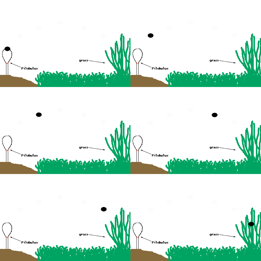
(b) Zoospores: They are thin-walled, motile spores formed in a zoosporangium. Example: In Pilobulus a sticky mass containing many spores is discharged as a single unit.
(c) Conidia: In some fungi the spores are not formed inside a sporangium. They are born freely on the tips of special branches called conidiophores. The spores thus formed are called as conidia. On the basis of development, two types of conidia are recognised namely thallospores and blastospores or true conidia.
(iii) Sexual reproduction:
With the exception of Deuteromycotina (Fungi imperfecti), the sexual reproduction is found in all groups of fungi. During sexual reproduction the compatible nuclei show a specific behaviour which is responsible for the onset of three distinct mycelial phases. The three phases of nuclear behaviour are as under:
Plasmogamy : Fusion of two protoplasts.
Karyogamy : Fusion of two nuclei.
Meiosis : The reduction division.
These three events are responsible for the arrival of the following three mycelial phases:
Haplophase : As a result of meiosis the haploid (n) or haplophase mycelium is formed.
Dikaryotic phase : The plasmogamy results in the formation of dikaryotic mycelium (n + n).
Diplophase : As a result of karyogamy the diplophase mycelium (2n) is formed.
Clamp Connection
In Basidiomycotina, the dikaryotic cells divide by clamp connections. They were first observed by Hoffman, (1856) who named it as 'Schnallenzellen' (buckle-joints).
Heterothallism
Blakeslee, (1904) while working with Mucor sp. observed that in some species sexual union was possible between two hyphae of the same mycelium, in others it occured between two hyphae derived from 'different' spores. He called the former phenomenon as homothallism and the latter as heterothallism.
|
|
Classification of Fungi
Phycomycetes
-
Members of phycomycetes are found in aquatic habitats and on decaying wood in moist and damp places or as obligate parasites on plants.
-
The mycelium is aseptate and coenocytic,Asexual reproduction takes place by zoospores (motile) or by aplanospores (non-motile).
-
These spores are endogeneously produced in sporangium.
-
Zygospores are formed by fusion of two gametes. These gametes are -similar in morphology (isogamous) or dissimilar (anisogamous or oogamous). Examples: Mucor, Rhizopus and Albugo (the parasitic fungi on mustard).
Rhizopus/Mucor
-
They are cosmopolitan and saprophytic fungus, living on dead organic matter.
-
Rhizopus stolnifer occur very frequently on moist bread, hence commonly called black bread mold.
-
Mucor is called dung mold.
-
Both are called black mold or pin mold because of black coloured pin head like sporangia.
-
Besides, it appears in the form of white cottony growth on moist fresh, organic matter, jams, jellies, cheese, pickles, etc.
Albugo
-
Albugo is a member of phycomycetes.
-
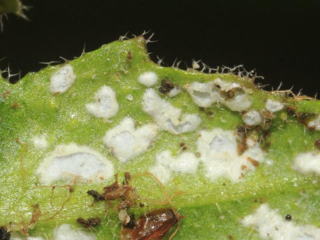
 It is an obligate parasite and grows in the intercellular spaces of host tissues. It is parasitic mainly on the members of families Cruciferae, Compositae, Amaranthaceae and Convolvulaceae, The disease caused by this fungus is known as white rust or white blisters.
 -
The most common and well known species is Albugo candida which attacks the embers of the mustard family (Cruciferae). It is commonly found on
-
Capsella bursa pastoris (Shepherd's purse), and occasionally on radish, mustard, cabbage, cauliflower, etc.
-
The reserve food is oil and Glycogen.
Ascomycetes
-
Commonly known as sac-fungi, the ascomycetes are unicellular, e.g., yeast (Sacharomyces) or multicellular, e.g., Penicillium.
-
They are saprophytic, decomposers, parasitic or coprophilous (growing on dung).
-
Mycelium is branched and septate.
-
The asexual spores are conidia produced exogenously on the special mycelium called conidiophores.
-
Sexual spores are called ascospores which are produced endogenously in sac like asci (singular ascus). These asci are arranged in different types of fruiting bodies called ascocarps.
-
Some examples are Aspergillus, Claviceps and Neurospora. Neurospora is used extensively in biochemical and genetic work. Many members like morels and buffles are edible and are-considered delicacies.
Yeast
-
Yeast was first described by Antony Von Leeuwenhoek in 1680.

-
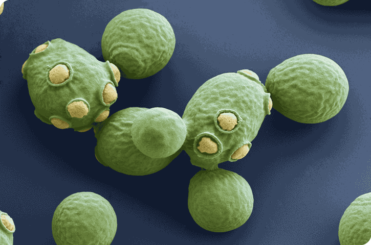
Yeast are nonmycelial or unicellular, which is very small and either spherical or oval in shape.
-
However, under favourable conditions they grow rapidly and form false mycelium or pseudomycelium.
-
Individual cells are colourless but the colonies may appear white, red, brown, creamy or yellow:
-
The single cell is about 10mm in diameter. It is enclosed in a delicate membrane which is not made up of fungal cellulose but is a mixture of two polysaccharides known as mannan and glycogen.
-
Reproduction: Yeast reproduces by vegetative or asexual and sexual methods.
(1) Vegetative reproduction: 'Yeast reproduce vegetatively either by budding or by fission.
(2) Sexual reproduction: Sexual reproduction in yeasts takes place during unfavourable conditions, particularly when there is less amount of food.The sex organs are not formed in yeasts 'and the sexual fusion occurs between the two haploid vegetative cells or two ascospores which behave as gametes. The two fusing gametes are haploid and may be isogamous or anisogamous. Such kind of sexual reproduction is called gametic copulation. It is the best example of hologamy i.e., the entire vegetative thallus is transformed into reproductive body. The sexual fusion leads to the formation of diploid zygote. The zygote behaves as an ascus and forms 4 - 8 haploid ascospores. These liberate and function as vegetative cells.
Basidiomycetes
-
Commonly known forms of basidiomycetes are mushrooms, bracket fungi or puffballs.
-
They grow in soil, on logs and tree stumps and in living plant bodies as parasites, e.g., rusts and smuts.

-
The mycelium is branched and septate.
-
The asexual spores are generally not found, but vegetative reproduction by fragmentation is common.
-
The sex organs are absent, but plasmogamy is brought about by fusion of two vegetative or somatic cells of different strains or genotypes.
-
The resultant structure is dikaryotic which ultimately gives rise to basidium.
-
Karyogamy and meiosis take place in the basidium producing four basidiospores.
-
The basidiospores are exogenously produced on the basidium.
-
The basidia are arranged in fruiting bodies called basidiocarps.
-
Some common members are Agaricus (mushroom), 'Ustilago (smut) and Puccinia (rust fungus).
Deuteromycetes 
-
Commonly known as imperfect fungi because only the asexual or vegetative phases of these fungi are known.
-
The deuteromycetes reproduce only by asexual spores known as conidia.
-
The mycelium is septate and branched.
-
Some members are saprophytes or parasites while a large number of them are decomposers of litter and help in mineral cycling. Examples: Alternaria, Colleiotrichum and Trichoderma.


Question 1: Embryophyta include:
(1) Algae
(2) Fungi
(3) Bryophyta
(4) All of these
Question 2: According to four kingdom system of Copeland, the fungi belong to kingdom –
(1) Protista
(2) Mychota
(3) Mycota
(4) Plantae
Question 3: Kingdom of unicellular eukaryotes –
(1) Monera
(2) Protista
(3) Fungi


| Q.1 | Q.2 | Q.3 |
| 3 | 1 | 2 |
Related Resources
-
Click here to refer the Useful Books of Biology for NEET (AIPMT)
-
Click here for Study Material on Kingdom Algae
To read more, Buy study materials of Biological Classification comprising study notes, revision notes, video lectures, previous year solved questions etc. Also browse for more study materials on Biology here.
Watch this Video for more reference
View courses by askIITians


Design classes One-on-One in your own way with Top IITians/Medical Professionals
Click Here Know More

Complete Self Study Package designed by Industry Leading Experts
Click Here Know More

Live 1-1 coding classes to unleash the Creator in your Child
Click Here Know More


